
Figure 3.
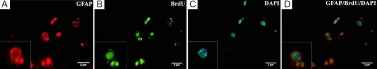
Expression of astrocyte marker glial fibrillary acidic protein (GFAP) in injured brain tissue after mesenchymal stem cell (MSC) transplantation.
(A) Some 5-bromo-2′-deoxyuridine (BrdU)-positive MSCs express GFAP. Labeled MSCs were indicated in red color. (B) BrdU labeled MSCs with fluorescein isothiocyanate (FITC)-labeled anti-mouse IgG antibody as secondary antibody were in green color. (C) 4′,6-Diamidino-2-phenylindole (DAPI)-labeled MSCs. DAPI indicated nuclei in blue color. (D) GFAP-BrdU-DAPI (× 1,000).