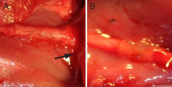
Figure 5.

Macroscale images for general observation of the sciatic nerve in the two groups at 12 weeks after repair.
(A) Adhesive group, (B) suture group. The repaired nerves showed good cooptation. The degree of tissue adhesion was mild, but slightly more clear in the distal end of the adhesive group (arrow). No neuro-ma formed in either group.
