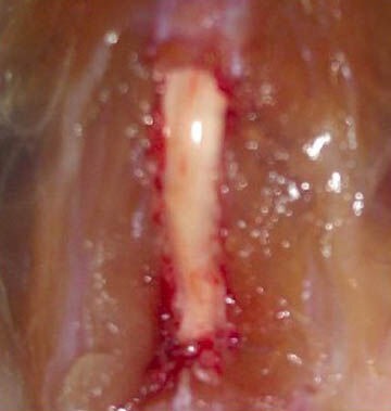
Figure 1.

Corresponding spinal segments associated with the sciatic nerve during modeling.
A 1.5 cm longitudinal incision was made on the posterior femur of the unilateral lower limb. The sciatic nerve trunk at the lower edge of the piriformis muscle was bluntly separated. The lamina was bitten to ex-pose the spinal segments associated with the sciatic nerve.
