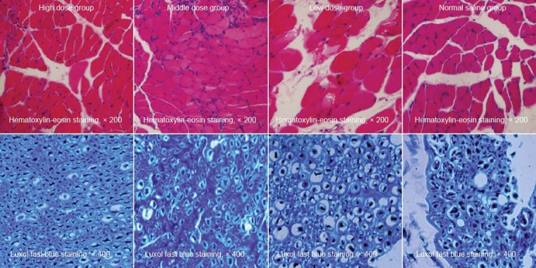
Figure 4.

Puerarin effects on the morphology of the triceps surae and nerve fibers of the spinal cord on the injured side at 8 weeks after sciatic nerve surgery.
The hematoxylin-eosin staining showed that muscle cells in the high and middle dose puerarin groups arranged regularly and tightly with a larger cross-sectional area of muscle fibers and smaller gaps. Luxol fast blue staining showed that the myelin sheaths arranged regularly with uniform thickness and clear outline and no proliferated fibrous connective tissues were found in the high and middle dose groups. While in the low dose group, the shape and thickness of the myelin sheaths were both irregular but still with a clear boundary, and fibrous connective tissues had prolifer-ated significantly; the normal saline group displayed disordered myelin sheaths and proliferated fibrous connective tissues.
