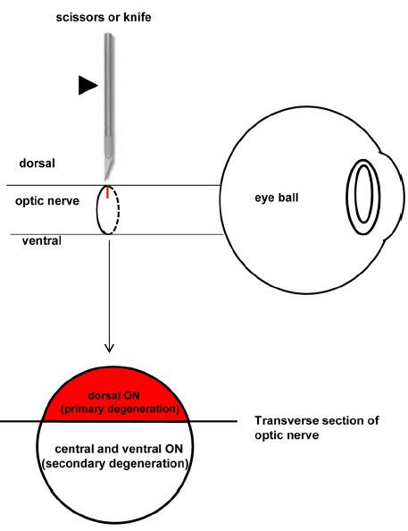
Figure 2.

Schematic diagram showing the surgery of partial optic nerve transection (PONT) and the location of primary and secondary degeneration in the optic nerve (ON).
The partial incision in the ON is achieved using a pair of scissors or a diamond knife (indicated by the arrowhead). The axons in the direct damaged sites (dorsal cut site of the ON in the transverse section, red color) will undergo primary degeneration and the axons in the indirect damaged sites (central and ventral areas of the ON in the transverse section, no color) will undergo secondary degeneration.
