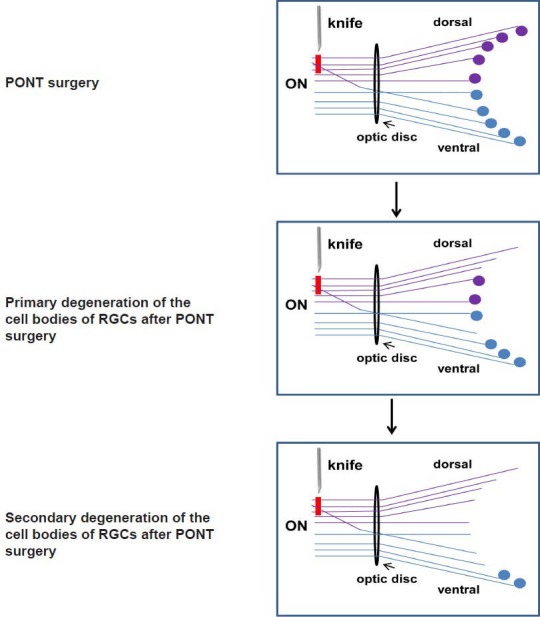
Figure 3.

The schematic diagram of the localization of both primary and secondary degeneration of the retinal ganglion cell (RGC) bodies in the retinas in Sprague-Dawley (SD) rats.
The lines indicate the axons in the optic nerve (ON) and the circles indicate the cell bodies of RGCs: the purple structures locate in the dorsal retina and dorsal ON and the blue structures locate in ventral retina and ventral ON. Red line indicates the axons transected after partial optic nerve transection (PONT) in the dorsal part of the ON.
