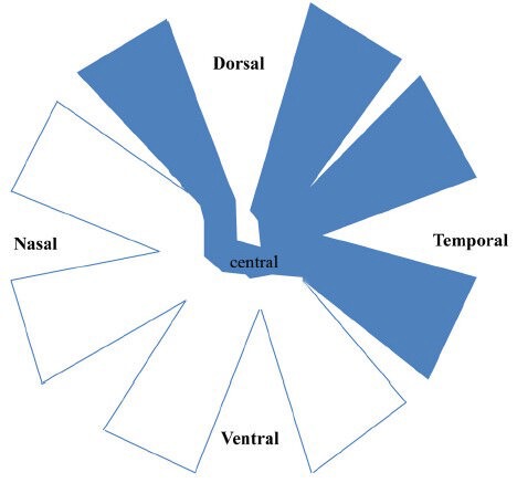
Figure 4.

The schematic diagram of the whole-mounted retinas of PVG hooded rats showing the location of the retinal ganglion cell (RGC) bodies whose axons were transected after partial optic nerve transection (PONT).
The retinas were divided into dorsal, ventral, nasal and temporal quarters in addition to the central area. The RGC bodies whose axons were transected after PONT located in the dorsal, temporal and central areas (indicated by blue color), but not in the nasal and ventral areas.
