
Figure 4.
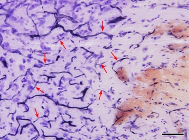
Sagittal section of spinal cord contusion (hematoxylin staining).
Red arrows, obstructed blood vessels. From right to left, reddish bleeding site of injury; pale ischemic zone with most of the neurons disappeared; an ischemic zone in which the morphology of most of the neurons remains largely normal. Bar = 100 μm.