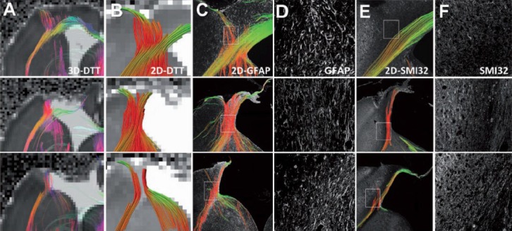
Figure 3.

Comparison of diffusion tensor tractography and histology-derived tractography demonstrates that the spurious fibers can be caused by reactive astrogliosis.
Three representative controlled cortical impact animals are shown. (A) Ex vivo 3D diffusion tensor tractography (DTT) maps depict tracts propagating in and along the lesion periphery similar to those observed in vivo. (B) 2D diffusion tensor tractography was subsequently performed to allow direct comparison to the 2D histology-derived tractography. (C) 2D tractography maps from the glial fibrillary acidic protein (GFAP)-stained sections revealed similarities to the DTI-derived maps near the lesion border. (D) The coherent orientation of astrocytes is shown on confocal images from selected regions. (E) 2D tractography maps from the Sternberger monoclonal antibody (SMI) 32-stained sections revealed fewer, if any, tracts propagating into the cortex near the lesion periphery. (F) A loss of structural integrity is noted for both the injured white matter and grey matter along the lesion border. These three examples demonstrate spurious WM tracts that are actually caused by reactive astrogliosis instead of fiber reorganization during brain plasticity. Image reproduced with permission from Budde et al. (2011) and Brain journal.
