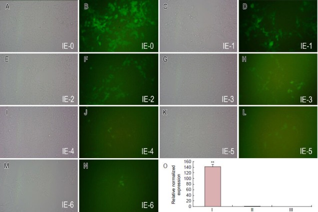
Figure 2.

miR-124 expression in lentiviral vector-infected BMSCs.
(A–N) In the 293FT cells transfected with the first dilution of the virus, there were numerous GFP-positive cells, as shown in Figure 4B. The num-ber of GFP-positive cells decreased with increasing dilution, until the seventh dilution, where three GFP-positive cells could be observed, as shown in Figure 4N. (O) The differences in expression of miR-124 were compared among miR-124+ (pLVX-EN-rno-miR124-transfected; I), miR-124– (pLVX-EN-rno-transfected; II) and control (untransfected; III) cells by RT-PCR. Bars represent mean ± SD. **P < 0.01, vs. the other two groups (Student's t-test) (n = 3). BMSCs: Bone marrow-derived mesenchymal stem cells; GFP: green fluorescent protein. IE-0–IE-6: Seven dilutions.
