
Figure 3.
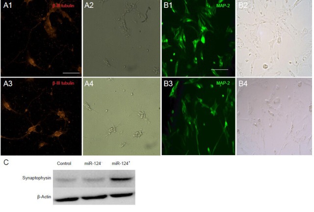
Neural differentiation of BMSCs on the 6th day of in vitro transfection.
(A, B) Immunofluorescence for the neuronal markers β-III tubulin (A1 and A3; red) and MAP-2 (B1 and B3; green) in BMSCs transfected with pLVX-EN-rno-miR124 (miR-124+) after 6 days of in vitro differentiation. Scale bars: 100 μm. BMSCs developed dendrites and neurites, similar to neurons (A2, A4, B2, B4). Scale bars: 100 μm. (C) Western blotting was used to detect the expression of synaptophysin (38 kDa). BMSCs: Bone marrow-derived mesenchymal stem cells; MAP-2: microtubule-associated protein-2.