
Figure 1.
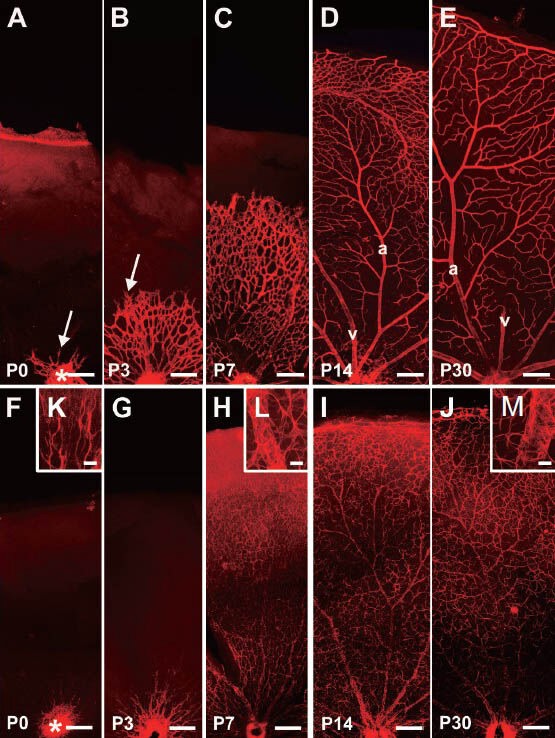
The retinal superficial vasculature system (red) and astrocytic network (red) of mice at different ages by Alex Fluro 568 immunofluorescentstaining.
(A–E) The retinal superficial vasculature system with collagen IV antibody immunofluorescence staining. The mouse retinal superficial vasculature radiates out from the optic disc (*)(A, B), eventually forming the mature vasculature with differing branching orders of arterioles and veins that cover the entire retina (C–E). The vascular buds, which are believed to be key germinal apparatuses for vasculogenesis, are marked with arrows. From P7, arteries and veins could be recognized. Usually, the arteries give rise to several large vessels into the retina; however, veins connected with the capillaries directly. In the photos, the arteries and veins are marked “a” and “v”, respectively. (F–M) The development of astrocytes at various ages with anti-glial fibrillary acidic protein antibody (GFAP) immunofluorescence staining. The immature retinal astrocytes appear from the optic disc (*) (F, G, K), eventually forming the astrocytic network in the entire retina (H–J). The processes of astrocytes contact each other to form the glial network, and many of them envelop vessels to participate in the formation of the blood-retinal barrier (H–J, L, M). Scale bars: A–J: 200 μm; K–M: 25 μm. P: Postnatal days.