Figure 2.

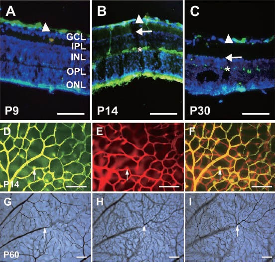
The vasculature in various layers of whole-mount retinas and retinal cross-sections by Alex Fluor 568 (red) and 488 (green) immunofluorescence staining and gelatin-ink (black) perfusion.
(A–C) The vascular distribution in retinal cross-sections with collagen IV immunofluorescence staining (green) and DAPI counterstaining cell nucleus (blue). The superficial vessels (arrowheads) were fully formed at P9, and were located in the inner surface of the ganglion cell layer. The deep vasculature formed at the inner edge (arrows) of the inner nuclear layer at first, and then invaded the outer edge (asterisks) of the inner nuclear layer at P14. The outer nuclear layer (ONL), outer plexiform layer (OPL) and inner plexiform layer (IPL) are marked as well. (D–F) The vascular distribution in a whole-mount retina at P14 (collagen IV immunofluorescence staining). The superficial vasculature at the inner surface of the ganglion cell layer (green) has been transformed into a green color using Photoshop software, and is shown in (D). The deep vasculature at the outer edge of the inner nuclear layer (red) is shown in (E). (F) is the merge of the images shown in D and E. The arrows indicate the branch into the deep vasculature. (G–I) The retinal vasculature in various layers visualized with ink staining at P60. The superficial vasculature (G) and deep vasculature at the inner edge (H) and outer edge (I) of the inner nuclear layer were visualized using different microscope focuses. The arrows point to a main vessel in the superficial vasculature, which branches into the deep vessels. Scale bars: 100 μm. P: Postnatal days.
