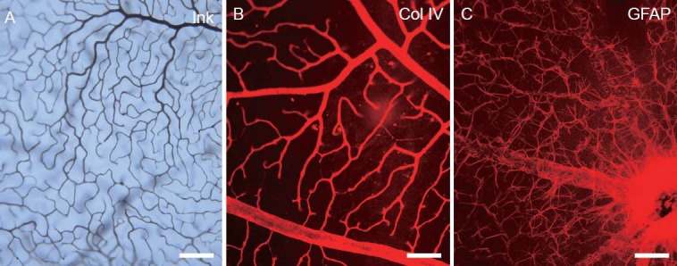
Figure 3.

The structure of the blood-retinal barrier of adult mice under light microscopy by gelatin-ink (black) perfusion, collagen IV and glial fibrillary acidic protein secondary antibody Alex Fluor 568 (red) immunofluorescence staining.
(A) The blood vessel lumen and endothelium is shown by gelatin-ink perfusion under a light microscope. (B) The basal lamina of the vessel is shown by anti-collagen (Col) IV immunofluorescence staining under a light microscope. (C) The astrocytic processes enveloping the retinal vessels are shown by anti-glial fibrillary acidic protein (GFAP) immunofluorescence staining under a light microscope. Scale bars: 100 μm.
