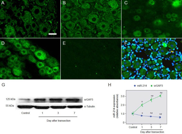
Figure 5.

Comparison of changes in srGAP3 and miR-214 levels in dorsal root ganglion neurons.
(A–F) srGAP3 detected by immunohistochemistry (scale bar: 20 μm). The level of srGAP3 was significantly upregulated in primary dorsal root ganglion neurons at 1 day (B), 3 days (C) and 7 days (D) after sciatic nerve transection, compared with levels in control dorsal root ganglion neu-rons (A). There was no specific labeling of srGAP3 in negative control sections (E). Overlays of srGAP3 (green) and DAPI (blue) staining showed cytoplasmic labeling of srGAP3 (F). (G) Western blot analysis of srGAP3 protein levels in control dorsal root ganglia and ipsilateral dorsal root ganglia at 1, 3 and 7 days after sciatic nerve transection. (H) Variation trends of miR-214 and srGAP3 levels in dorsal root ganglion neurons. Mean absorbance values were normalized using values for α-tubulin. The results are expressed as mean ± SEM. The quotient of the mean absorbance value/mean absorbance of each group (n = 6) and that of the control dorsal root ganglion neurons was calculated as a relative value. *P < 0.05, vs. control dorsal root ganglia; #P < 0.05, vs. 1 day after sciatic nerve transection using one-way analysis of variance and Dunnett's t-tests. srGAP3: Slit-Robo GTPase-activating protein 3; miR: microRNA.
