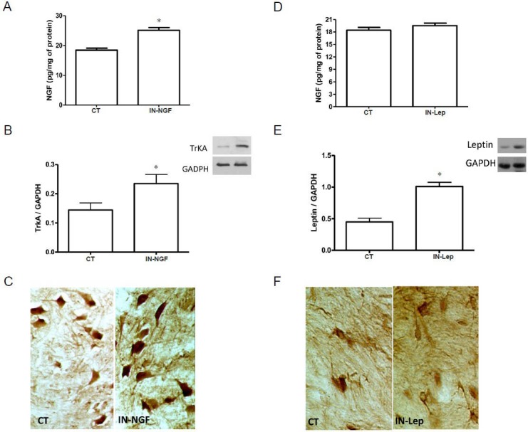
Figure 1.

Effects of IN-NGF or IN-Lep on spinal NGF in healthy rats.
(A) Levels of NGF in the spinal cord of rats 24 hours after receiving a single IN administration of vehicle (CT) or NGF (IN-NGF). The increase of NGF in the NGF-treated rats was revealed by ELISA. *P < 0.05, vs. CT. (B) Expression of TrkA in the spinal cord of rats 24 hours after receiving a single IN administration of vehicle (CT) or NGF (IN-NGF). A representative western blot is presented, together with data from densitometry analysis of five separate gel/blot runs (n = 5). As depicted in the panel, TrkA tissue content is increased in the IN-NGF-treated rats. Data are presented as arbitrary unit of grey levels after normalization with GAPDH band integrated optical density. *P < 0.05, vs. CT. (C) Immunohistochemical localization of TrkA in the spinal cord of rats 24 hours after receiving a single IN administration of vehicle (CT) or NGF (IN-NGF). The picture shows the increase of TrkA immunoreactivity in spinal cord neurons in NGF-treated rats. (D) Levels of NGF in the spinal cord of rats 24 hours af-ter receiving a single IN administration of vehicle (CT) or leptin (IN-Lep). As revealed by ELISA, spinal NGF levels were unaffected by IN-Lep. (E, F) Leptin levels in the spinal cord after a single IN administration of vehicle (CT) or leptin (Lep). A representative western blot is presented in panel E, together with data from densitometry analysis of five separate gel/blot runs (n = 5). As depicted in the panel, Lep tissue content is increased in the IN-Lep-treated rats. Data are presented as arbitrary unit of grey levels after normalization with GAPDH band integrated optical density. *P < 0.05, vs. CT. The immunohistochemical localization of leptin in the spinal cord (panel F) revealed that the protein immunoreactivity increases after IN-Lep administration. IN: Intranasal; IN-NGF: intranasal administration of nerve growth factor; IN-Lep: intranasal administration of leptin.
