Figure 6.

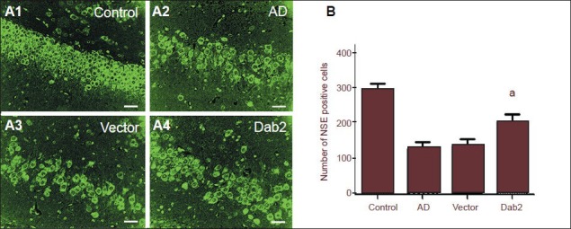
Effect of Dab2 overexpression on hippocampal neurons in Alzheimer's disease (AD) mice.
(A1–A4) By fluorescence microscopy, neuron-specific enolase (NSE)-positive cells exhibited green fluorescence in the cytoplasm. Hippocampal neurons are arranged compactly and orderly in the control group. Hippocampal neurons were reduced in number and arranged in a disorderly fashion in the AD and vector groups. The level of reduction in hippocampal neurons was lower in the Dab2 group compared with the AD group. Scale bars: 10 μm. (B) The number of hippocampal NSE-positive cells in the Dab2 group was significantly higher than the numbers in the vector and AD groups (aP < 0.05). One-way analysis of variance and the SNK-q test were used to assess the significance of differences between groups. Data are represented as mean ± SEM from at least three independent experiments.
