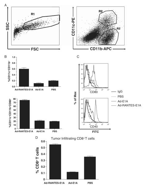
FIGURE 2.
RANTES expression in the tumor site attracts the antigen-presenting cells into the tumor mass. A, 5 × 10⁵ JC tumor cells were subcutaneously inoculated and the established tumors were injected with 1010 i.f.u. of Ad-RANTES-E1A, Ad-E1A, or PBS when they reached 5 to 7 mm in diameter. Tumors were established in 3 mice for each treatment group and processed separately. Forty-eight hours later, tumors were resected and total tumor single-cell suspensions (side and forward) scatter analyzed by flow cytometry depicted on left panel, APCs (gated as R1) were stained with antimouse CD11c, CD11b, and CD80 mAbs (right panel, flow cytometry results for CD11c and CD11b expression). DCs were gated as CD11c+ CD11b+ cells (gate R2); macrophages were gated as CD11c− CD11b+ (gate R3). Mean percentage of DCs (B, upper panel). Maturation status of DCs was determined by staining with monoclonal antibodies to mouse CD80 and CD40 [B (lower panel) and C]. D, Percentage of tumor infiltrating CD8+ T cells. Data presented for 3 mice. DC indicates dendritic cell; mAbs, monoclonal antibodies; PBS, phosphate buffered saline; RANTES, regulated upon activation, normally T expressed, and presumably secreted.

