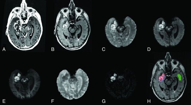
Fig 1.
Anatomic T1-weighted postcontrast (A) and T2-weighted FLAIR (B) in an 84-year-old man with glioblastoma (patient 3). DWI images at b = 500 (C), b = 1500 (D), and b = 4000 (E); ADC (F); and an RSI-CM are shown (G). VOIs for tumor (red), peritumoral edema (blue), and normal-appearing WM (green) used for quantitative analysis are shown in H.

