Fig. 3.
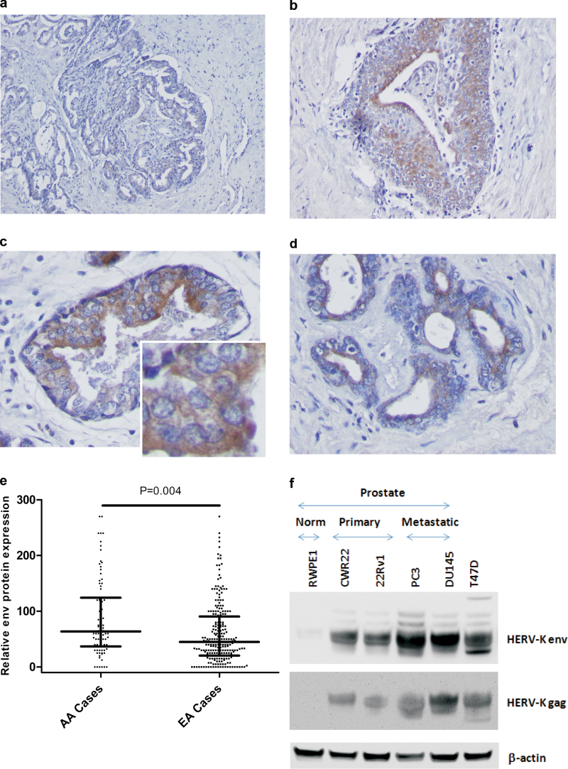
HERV-K expression in human prostate adenocarcinomas. Shown is IHC analysis of four invasive adenocarcinomas for expression of HERV-K envelope (env) protein using the monoclonal anti-envelope antibody, 6H5. (a) Scattered positivity for HERV-K env expression in the tumor epithelium, showing a low to moderate antigen expression as indicated by the brown chromogen deposits. (b–d) Locally intensive staining for env expression in the tumor epithelium. Staining shows a cytosolic to membrane distribution with a more intensive staining of cancer cells toward the luminal side of the cancerous gland (c and d). a and b: Magnification: ×100. c and d: Magnification: ×400. Inset: higher resolution image for the env protein-positive tumor epithelium. Counterstain: Hematoxylin. (e) Immunostaining for env protein in human prostate tumors using a tissue microarray that included tumors from both African-American (n = 105) and European-American patients (n = 272). On average, tumors from African-American patients showed a higher expression of the HERV-K env protein than tumors from European-Americans (Student’s t-test, P < 0.001). (f) HERV-K gag and env protein expression were detected in human prostate cancer cell lines (CWR22, 22Rv1, PC-3 and DU145) but were not detected in the non-tumorigenic human prostate cell line, RWPE1. An extract from the HERV-K-positive T47D human breast cancer cell line were included as a positive control.

