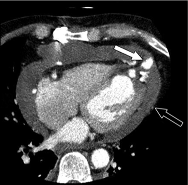
Fig. 2.

45-year-old man who underwent cardiac CT angiography 6 days after coronary bypass grafting surgery. Axial CT image shows anterior ventricular pseudoaneurysm (white arrow) and large hemopericardium (open arrow). Patient had good recovery after surgery.
