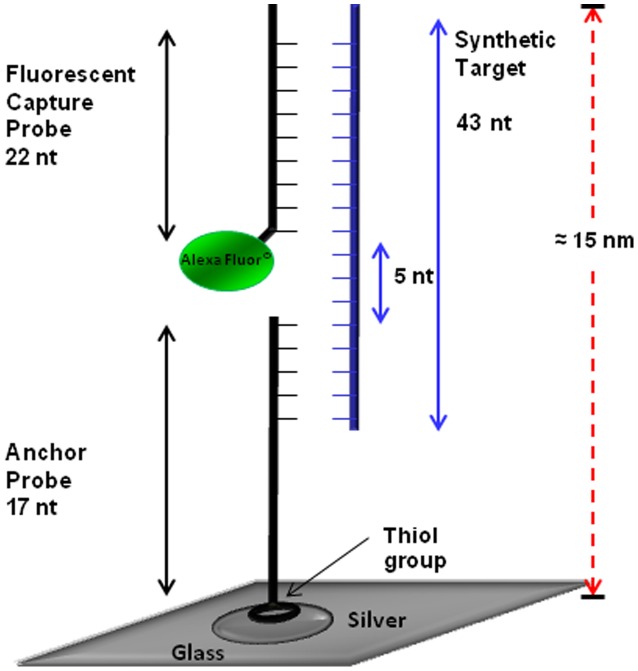
Figure 2. Schematic configuration of 3 piece DNA detection assay.
This configuration was used for detection of both toxin A and toxin B probes. The anchor probe (17 oligonucleotides) was anchored to the SiFs by addition of the thiol group. The fluorescent probe (22 oligonucleotides) was attached to an Alexa at the 3′ end. Upon hybridization with target DNA, the 3 piece assay is formed, and the fluorophore labeled probe is plasmon enhanced.

