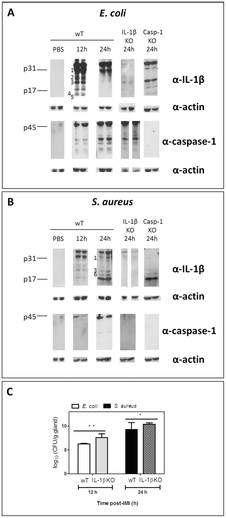
Figure 3. Differential mammary IL-1beta fragmentation post-IMI with E. coli versus S. aureus and effect on bacterial growth.
(A) Cleavage of IL-1beta in the mammary gland post-IMI with E. coli shows a fast, transient IL-1beta maturation with six IL-1beta fragments at 12h i.e. ±30 kDa, 25 kDa, ±20 kDa, ±17 kDa, ±15 kDa and ±10 kDa (fragments 1, 2, 3, p17, 4 and 5, respectively). At 24 h (only fragment 1 and p31 proform) is seen. In IL-1beta KO and in sham-inoculated (PBS) mice no fragments or p31 proform are detected. Despite clear procaspase-1 maturation, the early complex IL-1beta pattern is not the result of caspase-1 cleavage as the latter only occurs extensively at 24 h and as cleavage of pro-IL-1beta still occurs in caspase-1 KO glands (right Western blot images). No caspase-1 maturation was detected in caspase-1 KO or in sham-inoculated mammary glands. (B) Cleavage of IL-1beta in the mammary gland post-IMI with S. aureus shows a slower IL-1beta maturation with four IL-1beta fragments at 24 h i.e. ±30 kDa, ±20 kDa, ±18 kDa and ±17 kDa (fragments 1, 3, 6 and p17, respectively). At 12 h (only fragment 1 i.e the preform p31, 2, 3,and p17). The late IL-1beta maturation is not the result of caspase-1 cleavage as IL-1beta cleavage still occurs in caspase-1 KO glands (strong p17 fragment albeit in the absence of the p31 proform). Both procaspase-1 and its cleavage were low, respectively absent in IL-1beta KO glands (right Western blot images). No IL-1beta or caspase-1 was detected in sham-inoculated mammary glands of wT mice. (C) ProIL-1beta fragmentation affects bacterial growth as in IL-1beta KO mammary glands on time points of interest (12 h for E. coli, 24 h for S. aureus) CFU counts are significantly higher for both pathogens than in wT mammary glands, especially for E. coli (P<0.01 *, P<0.001**). DL = detection limit.

