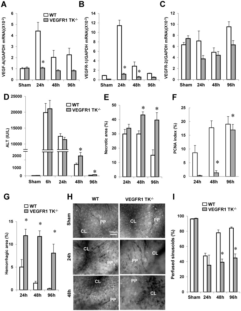
Figure 1. Delayed liver repair and sinusoidal reconstruction after hepatic I/R in VEGFR1 TK-/- mice.
Changes in VEGF-A (A), VEGFR1 (B), and VEGFR2 (C) mRNA levels in livers from WT mice and VEGFR1 TK-/- mice after hepatic I/R, and changes in ALT levels (D), the area of hepatic necrosis (E), the PCNA index (F), and the hemorrhagic area (G). Representative in vivo microscopic images showing the uptake of acetylated LDL (white dots) at 24 h and 48 h (H). PP, periportal region; CL, centrilobular region. Sinusoidal perfusion after hepatic I/R (I). Data are expressed as the mean ± SEM from five to six mice per group. *p<0.05 vs. WT mice.

