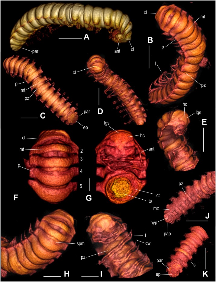
Figure 3. Maatidesmus paachtun nov. gen. and sp., holotype, 3D micro-CT reconstruction.
(A–D): General view, scale bar 5 mm. E) Head, collum and first segments in lateral view, scale bar 5 mm. (F) Dorsal view of first segments, scale bar 2 mm. (G) Ventral view of head with a cross section of first segments, scale bar 2 mm. (H) Collum, metaterga and paranota in dorsal view, scale bar 3 mm. (I) Head and legs in dorsal view, scale bar 3 mm. (J) Last segments in ventral and dorsal view (K), respectively, scale bar 5 mm. All 3D images are expressed in virtual colors. See anatomical abbreviations in the main text.

