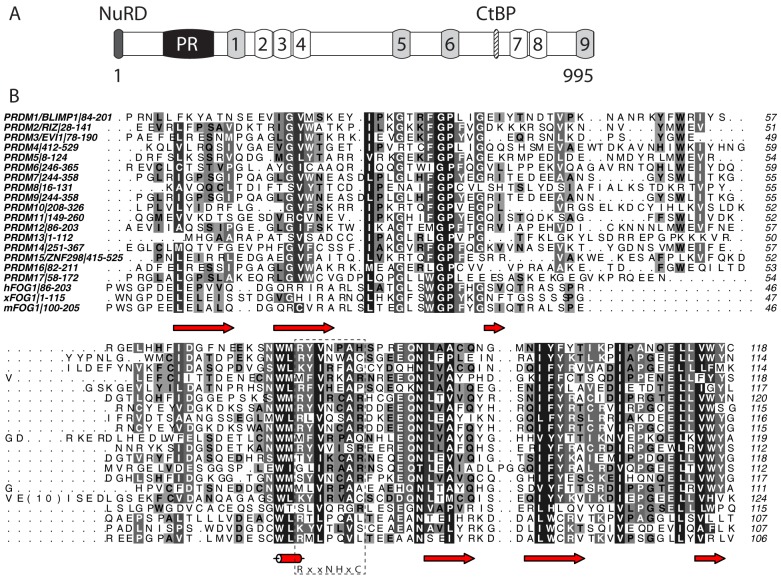
Figure 1. Domain structure and sequence analysis of FOG1.
(A) Domain structure of murine FOG1. C2HC type zinc fingers are light grey; C2H2 fingers are unshaded. The binding sites for CtBP and the NuRD complex are indicated, as is the newly identified PR domain. (B) Sequence alignment of murine FOG(100–205) with human and Xenopus laevis FOG1 and with all human PR domains. Colouring indicates conservation at four different levels. The essential catalytic consensus motif found in SET domains is shown below the alignment and indicated with a dashed box. Secondary structure elements in FOG-PR are indicated below the alignment. Alignment was carried out using CLUSTAL OMEGA [49] and the diagram made using ALINE [50].

