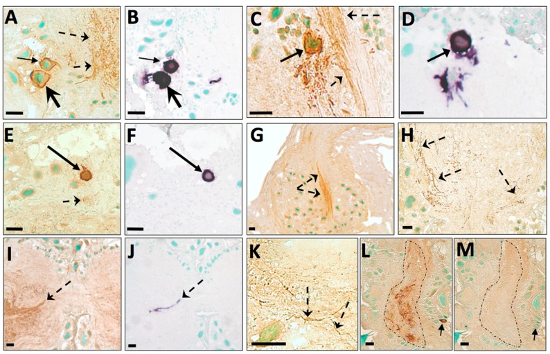
Figure 3. ISH and ICC of A. californica CNS.
Representative photomicrographs of ISH (purple; B, D, F, J) or ICC (brown; A, C, E, G, H, I, K, L, M) staining in the CNS of A. californica, including abdominal (A, B, L, M), buccal (K), cerebral (C, D, I, J), pleural (E, F), and pedal (H) ganglia, and bag cell neurons (G). Preadsorption with ap-AKH (M) completely abolished ap-AKH-ir signal compared to an adjacent section (L). Green = methyl green nuclear counterstain. Solid arrows denote ap-AKH-ir or ap-AKH transcript-positive neuronal cell bodies; arrow pairs of the same shape in panels A/B, C/D, E/F, and L/M point to identical neurons in adjacent sections. Dashed arrows in panels A, C, E, G, H, I, and K point to ap-AKH-ir fibers in the neuropil regions of ganglia. Dashed arrow in panel J points to ap-AKH transcript-positive fibers. Dashed outline in panel L surrounds ap-AKH-ir fibers in the neuropil region of the abdominal ganglia, and in M surrounds the same region of neuropil devoid of signals. Scale bars = 50 µm.

