Figure 2.
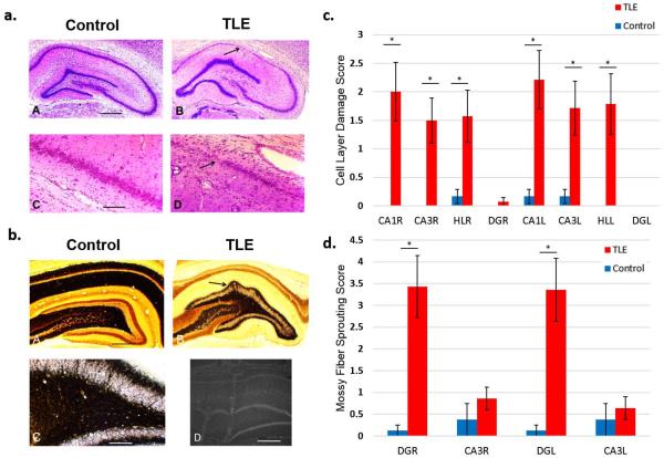
TLE rats show extensive cell loss and damage in the CA1 and CA3 layers as well as the Hilus (HL) and the Dentate Gyrus (DG) of the hippocampus and mossy fiber sprouting in DG. (a) Cresyl violet staining images. Calibration bar = 1 mm (a), 400 μm (b). Example images of hippocampal regions from TLE and control rats where cell layer damage was assessed. (b) TIMM staining images for assessment of mossy fiber sprouting. Example of mossy fiber sprouting in the hippocampal regions of TLE and control rats (A–C). Electrode placement was confirmed by dipping recording electrodes into fluorescent dye DiI (D). (c) Histological assessment of cell layer damage (Table 1) in the hippocampal regions using Cresyl violet staining. Bilateral cell layer damage in the CA1, CA3, DG, and HL was observed in TLE rats. (d) Histological assessment of mossy fiber sprouting (Table 2) in DG and CA3 regions of the hippocampus using TIMM staining. Bilateral mossy fiber sprouting was present in the DG but not in the CA3 layers. TLE rats were observed to have severe bilateral cell loss in the CA1, CA3 and HL regions as well as extensive bilateral mossy fiber sprouting in the DG. * P <0.001

