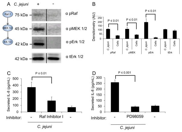
Fig. 2. C. jejuni activates Erk 1/2 by the canonical MAPK activation pathway.
Panels: (A) Immunoblot analysis of INT 407 cells infected with C. jejuni following a 30 min incubation. The level of phosphorylated c-Raf, MEK 1/2 and Erk 1/2 were determined by immunoblot with phospho-specific monoclonal antibodies for each protein. Total Erk 1/2 (tErk) served as a loading control. The images shown are a composite of different sections of the same immunoblot. (B) Densitometric analysis of phosphorylated Raf/MEK/Erk from three samples. (C) INT 407 cells were treated with Raf Inhibitor I and IL-8 secretion was determined by ELISA 24 hr following infection with C. jejuni. Uninfected cells served as a negative control. (D) INT 407 cells were treated with the Erk 1/2 activation inhibitor PD98059, infected with C. jejuni, and the level of IL-8 in supernatants were measured by ELISA. Vehicle treated cells infected with C. jejuni served as a positive control and uninfected cells served as a negative control.

