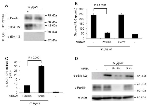
Fig. 6. Erk 1/2 is recruited to and activated at paxillin in response to C. jejuni infection.
Panels: (A) INT 407 cells were infected with C. jejuni and incubated for 40 min. Uninfected cells served as a negative control. Paxillin was immunoprecipitated from cell lysates with anti-paxillin antibody. The cell lysates were analyzed by immunoblot and probed with antibody against total Erk 1/2. Immunoprecipitations using antibodies against IgG were used to examine nonspecific interactions. (B) INT 407 cells were treated with siRNA specific to paxillin and infected with C. jejuni. Lysates were collected and immunoblots probed with antibodies directed against phospho Erk 1/2 (pErk), with total Erk 1/2 (tErk) serving as a loading control. The efficiency of paxillin knockdown using siRNA treatment was tested by immunoblot analysis. (C) INT 407 cells were transfected with siRNA specific to paxillin or a scambled (Scrm) control and infected with C. jejuni. The level of IL-8 gene expression was measured by RT-PCR following a 6 hr infection. IL-8 expression was normalized to GAPDH transcript levels. Uninfected cells served as negative control. (D) ELISA quantification of IL-8 secretion by INT 407 cells treated with siRNA specific to paxillin or scrambled siRNA following incubation with C. jejuni. Uninfected cells served as a negative control.

