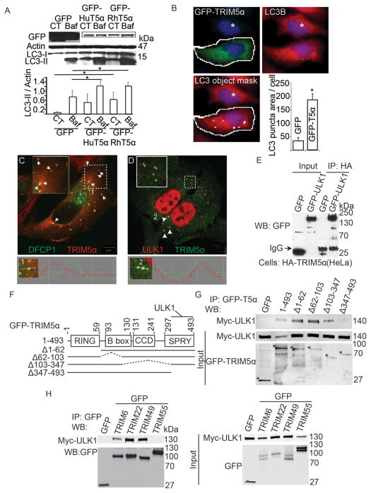
Figure 3. TRIM5α and additional TRIMs interact with ULK1.
(A) Top, 293T cells were transfected with GFP-tagged human (HuT5α) or rhesus (RhT5α) TRIM5α or GFP alone and treated or not with bafilomycin A1 (Baf) and levels of LC3B and actin were assayed by immunoblot. Bottom, quantitation of LC3B-II/actin ratios. CT, control without Baf. (B) High content analysis of endogenous LC3 puncta in HeLa cells (full media) transfected with GFP or GFP-TRIM5α. Object masks: White contour line, gating for primary objects (GFP-positive cells). White internal small object masks, LC3B puncta. White asterisk, GFP-negative cell (manually entered; excluded from analysis by iDEV software). Graph, quantification of LC3B puncta area per green fluorescent (GFP+ or GFP-TRIM5α+) cell. Data, means ± SE, n ≥ 3 experiments, *, P bold> 0.05 (t test). (C) Confocal microscopy of HeLa cells stably expressing HA-TRIM5α and transiently expressing GFP-DFCP1. Numeral 1, a punctum displayed in line tracing profile below. (D) Confocal microscopy analysis of HA-tagged TRIM5α (green) and endogenous ULK1 (red) in HeLa cells. Numeral 2, two puncta displayed in line tracing profile below. (E) Lysates from HeLa cells stably expressing HA-tagged TRIM5α and transiently over-expressing either GFP-ULK1 or GFP alone were subjected to immunoprecipitation with anti-HA and immunoblots probed with anti-GFP. (F) Domain organization of TRIM5α and deletion constructs used in mapping experiments. (G) Co-immunoprecipitation analysis of full-length or deletion variants of TRIM5α (as GFP fusions; asterisks denote fusion products on the bottom blot) with Myc-ULK1 transiently expressed in 293T cells. (H) Co-immunoprecipitation analysis of interactions between indicated TRIMs (as GFP fusions) and Myc-ULK1 in 293T cell extracts. See also Figure S3.

