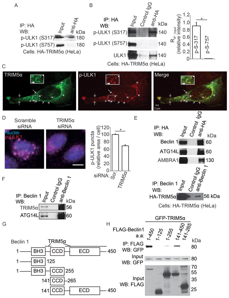
Figure 4. TRIM5α interacts with activated ULK1 and with Beclin 1.
(A) Lysates from HeLa cells stably expressing HA-TRIM5α and transiently transfected with GFP-ULK1 were immunoprecipitated with anti-HA and blots probed with antibodies against Ser-317 or Ser-757 phospho-ULK1. (B) Lysates from HeLa cells stably expressing HA-TRIM5α were immunoprecipitated with anti-HA and blots probed as in A for endogenous ULK1. Graph, ratio of immunoprecipitated phospho-ULK1 to phospho-ULK1 in the input. (C) Confocal microscopy of cells stably expressing HA-tagged TRIM5α (green) and stained to detect p-ULK1 (p-Ser 317; red). Arrows, overlaps between p-ULK1 and HA-TRIM5α. (D) High content analysis of endogenous p-ULK1 (p-Ser 317; red) in control HeLa cells or cells subjected to TRIM5α knock-down. Blue, nuclei. Graph, area of phospho-ULK1 (Ser-317) puncta per cell. (E) Top, lysates from cells stably expressing HA-tagged TRIM5α were immunoprecipitated with anti-HA and immunoblots probed as indicated. Bottom, lysates as above were subjected to immunoprecipitated with anti-Beclin 1 and immunoblots probed for HA-TRIM5α. (F) Lysates from rhesus epithelial cells (FRhK4) were immunoprecipitated with anti-Beclin 1 and immunoblots probed with the indicated antisera. (G,H) Mapping of Beclin 1 regions interacting with TRIM5α. Schematic (G) of Beclin 1 constructs used in immunoprecipitation experiments in H. Lysates of 293T cells co-expressing GFP-tagged TRIM5α and the indicated FLAG-tagged Beclin 1 constructs in D were immunoprecipitated with anti-GFP and immunoblots probed as indicated. Data, means ± SE, n ≥ 3 experiments, *, P < 0.05 (t test). See also Figure S4.

