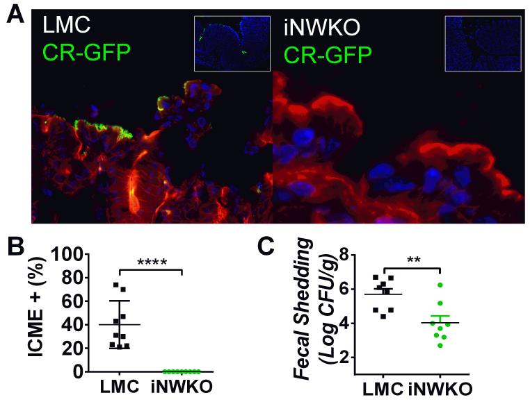
Figure 2. Intestinal N-WASP is required for colonization of mice by C. rodentium.
(A) Littermate control and iNWKO mice were infected with 5×10⁸ GFP-C. rodentium by oral gavage. At four days post-infection, after DAPI-staining (blue) to visualize intestinal cells and phalloidin-staining (red) to visualize the cell architecture, large numbers of adherent GFP-positive C. rodentium were identified in littermate control mice (left panel) but not iNWKO mice (right panel). Inset shows low power view of DAPI and GFP, indicating the absence of identifiable C. rodentium in iNWKO mice. (B) Intercrypt mucosal epithelium (ICME) with adherent GFP+ C. rodentium was quantified in littermate control and iNWKO mice at four days post-infection. Shown is the mean ± SD. Statistical significance was determined by one-way t-test and ****, p<0.0001. (C) Fecal shedding (±SEM) of iNWKO or littermate control mice at seven days post-infection by C. rodentium was determined by quantifying viable stool counts. Data reflect a compilation of two experiments, with a total of four mice per group per experiment. (**p<0.01; by an unpaired t-test).

