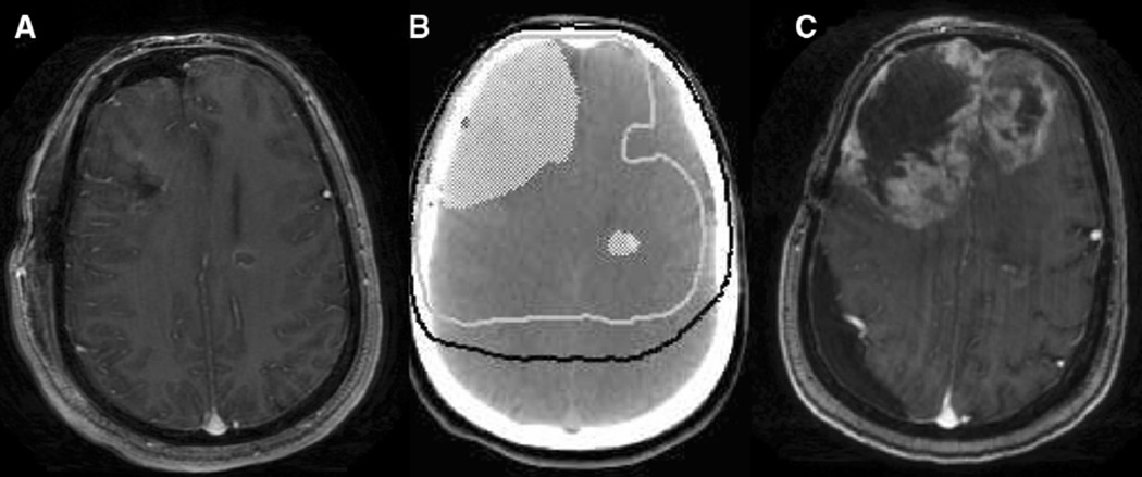
Fig. 1.
a T1 axial contrast-enhanced MRI done in the post-operative for multicentric GBM. There is a resection cavity in the right frontal lobe and a residual ring-enhancing lesion in the left frontal lobe. 1b Treatment planning axial non-contrast-enhanced CT demonstrating post-operative tumor volume in white colorwash, 60 Gy isodose line represented as white solid line, and 46 Gy isodose line represented as black solid line. 1c T1 axial contrast-enhanced MRI done at time of tumor progression showing tumor progression predominantly in the highest dose radiation volume

