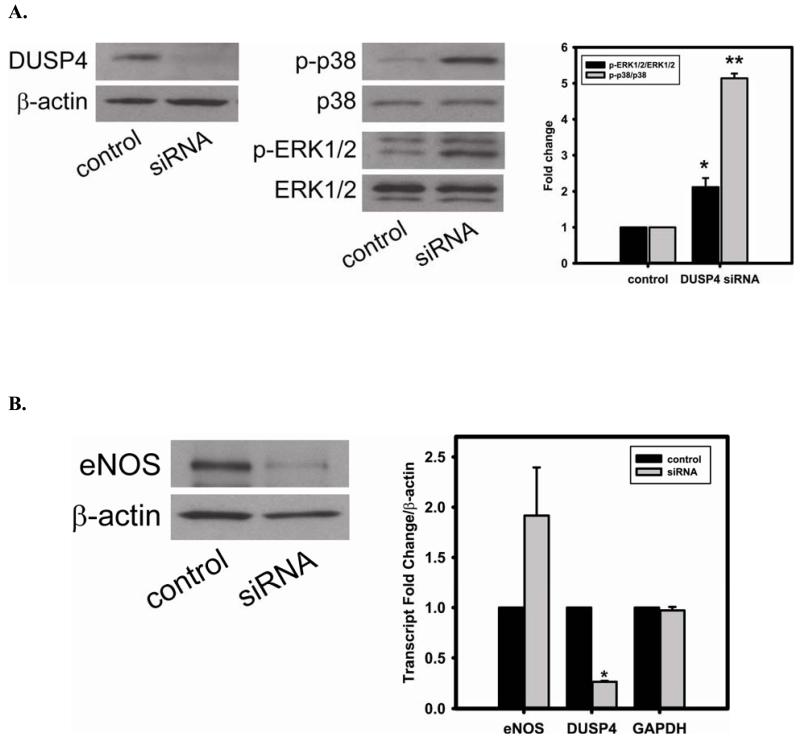
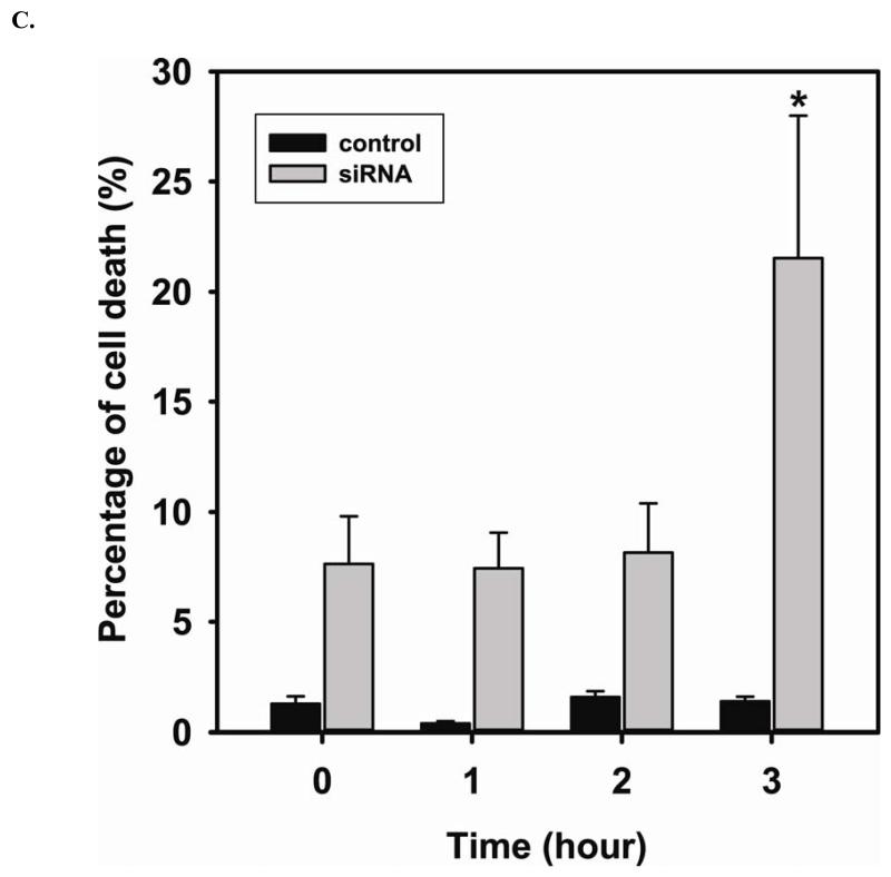
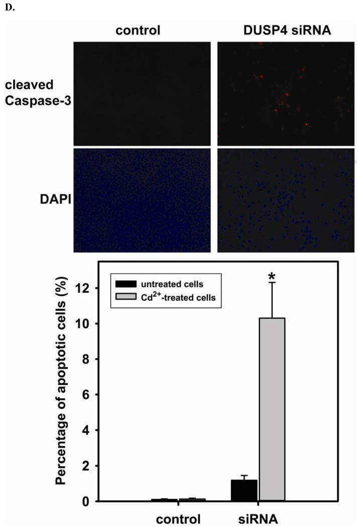
FIGURE 5. DUSP4-dependent eNOS expression, and the modulation of p38 and ERK1/2 signal cascades in RAECs.
A. DUSP4 gene silencing. Left: The efficiency of DUSP4 gene silencing is greater than 80% as determined by immunoblotting against DUSP4, with β-actin as a loading control. Middle: DUSP4 gene silencing contributes to the over-activation of both p38 (upper panel) and ERK1/2 (lower panel). Right: Fold change of the ratio of p-ERK1/2/ERK1/2 (2.12 ± 0.25 fold versus control; * P < 0.05) and p-p38/p38 (5.14 ± 0.13 fold versus control; ** P < 0.0001). B. DUSP4 gene silencing leads to a decrease in eNOS expression (0.55 ± 0.12 fold versus control; P < 0.05) via the translational modulation. Left: immunoblotting of eNOS and β-actin as a loading control. Right: Quantitative mRNA analysis of eNOS, DUSP4, and GAPDH using β-actin as an internal control. DUSP4 siRNA only affects DUSP4 gene expression (0.27 ± 0.01 fold versus control; * P < 0.0001), but not eNOS or GAPDH. C. Time course of cell death during 3 hr Cd2+ exposure. Rat endothelial cells with DUSP4 knockdown are more susceptible to Cd2+-induced death. Greater cell death is seen in cells with DUSP4 gene silencing compared to control cells after 3 hr Cd2+ exposure (21.54% ± 6.46% versus control; * P < 0.05). D. Immunostaining against cleaved caspase-3. Rat endothelial cells with DUSP4 knockdown are susceptible to Cd2+-induced apoptosis. More cleaved caspase-3 positive is seen in cells with DUSP4 gene silencing compared to control cells after 3 hr Cd2+ exposure (10.31% ± 2.01% versus control; * P < 0.05).


