Figure 2.
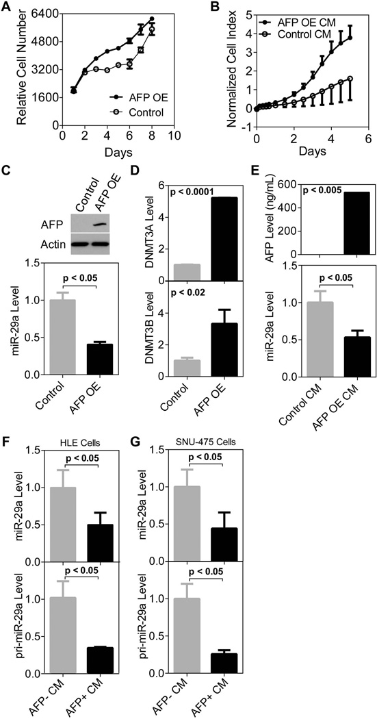
AFP transcriptionally regulates miR-29. A) Over expression of AFP significantly increases cell proliferation compared to control HLE cells after a 48hr transient transfection (p<0.0005 days 3–6 and p<0.005 days 7 and 8). Cell growth was monitored using a CalceinAM assay with 4 replicates per time point and error bars represent standard deviation. B) HLE cells in the presence of conditioned media taken from AFP over expressing HLE cells (AFP OE CM) proliferate faster than those in AFP− CM (taken from HLE cells transfected with an empty vector, Control CM). Cells were seeded on day 0, CM was applied on day 1 and cell growth was monitored by xCELLigence technology over a period of five days. There are four replicates per time point and error bars represent standard deviation. The growth rate of HLE cells growing in the presence of AFP is significantly faster on days 1–5 (p<0.05). C) Over expression of AFP in HLE cells leads to decreased mature miR-29a expression compared to control HLE cells (48 hour transient transfection). Top panel shows AFP protein expression by western blot. miR-29a expression was quantified using qRT-PCR in the bottom panel. D) DNMT3A and DNMT3B levels significantly increased after transient AFP over expression (48 hours) in HLE cells. E) Conditioned media taken from transfected cells was applied to HLE cells. AFP OE CM led to a significant decrease in mature miR-29a measured by qRT-PCR as compared to CM from control cells. ELISA data in the top panel shows that media taken from cells over expressing AFP has >500ng/ml of AFP present. F) AFP+ CM taken from HUH-7 cells led to a decrease in both the mature (top panel) and primary transcript (bottom panel) of miR-29a in HLE cells as measured by qRT-PCR. G) AFP+ CM from HUH-7 cells also lead to a decrease in miR-29a mature (top panel) and primary transcript (bottom panel) expression in SNU-475 cells.

