
Figure 2. Activity of CEl PKC-δ+ neurons is required for the influence of anorexigenic agents.
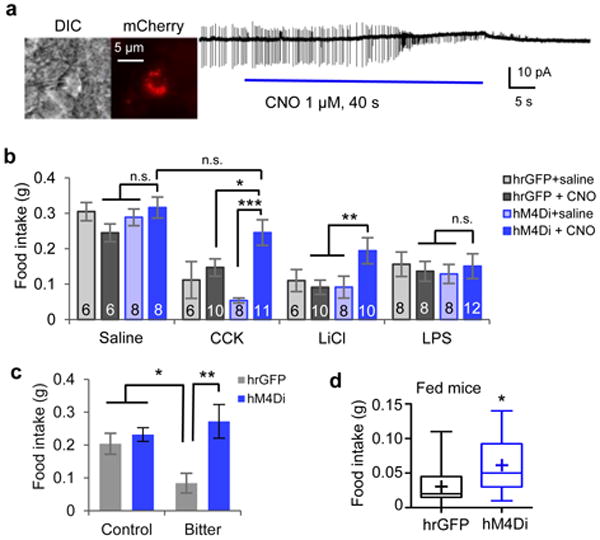
a. Cell-attached slice recording from a CEl PKC-δ+ neuron expressing hM4Di-mCherry that is silenced by bath application of CNO. b. Food intake in 24 h fasted animals after administration of different anorexigenic agents. The number of animals in each condition is indicated in the bars. Values are means ± s.e.m.. Two-way ANOVA with post-hoc Bonferroni t-tests showed a significant effect of CNO silencing, F(1, 24) = 0.363, p = 0.55 (saline); F(1, 31) = 11.7, p = 0.0018 (CCK); F(1, 30) = 4.50, p = 0.042 (LiCl); F(1, 32) = 0.001, p = 0.98 (LPS). c. Intake of control vs. quinine-laced food in 24 h fasted animals expressing hrGFP (n = 5 animals) or hM4Di (n = 5 animals), after intraperitoneal injection of CNO. Note that addition of quinine (bitter) inhibited food intake in controls. Values are means ± s.e.m.. Two-way ANOVA (F(1, 16) = 9.91, p = 0.0062) with post-hoc Bonferroni t-test showed a significant effect of silencing. d. Food intake in CNO-treated fed mice expressing hrGFP (n = 13 animals) or hM4Di (n = 14 animals) in CEl PKC-δ+ neurons. Box plots show mean (+), median, quartiles (boxes), and range (whiskers). Unpaired t-test, t(25) = 2.2, p = 0.045. n.s., not significant; * p < 0.05, ** p < 0.01.