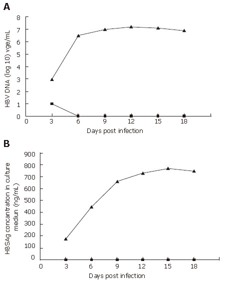
Figure 4.

The efficiency of PHH infected with wt HBV or rHBV. PHH were infected at a multiplicity of infection of 10 DNA-containing HBV particles/cell on d 1 post-seeding, HBV DNA (A), and HBSAg (B) secreted into cell culture medium were determined every 3 d after infection. The triangles denote PHH cells infected with wt HBV, and squares denote PHH cells infected with rHBV. In the supernatant from PHH infected with rHBV, no HBV DNA and HBsAg can be detected, meaning that all viral gene were knocked out.
