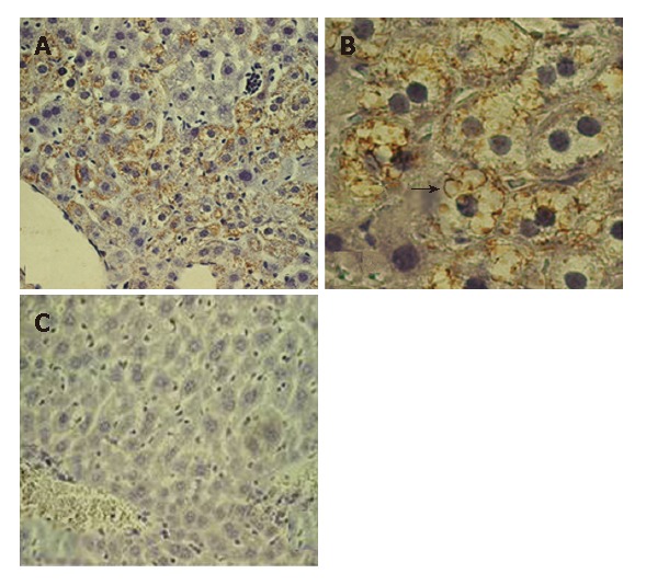
Figure 3.

Immunostaining of the female 2 mo old DTM liver (without Dox). A: The core positive hepatocytes are grouped near a central vein (× 20); B: The characteristic globular core expression is shown clearly (arrow, × 60); C: The sex-age matched DTM on Dox chow mouse liver served as negative control.
