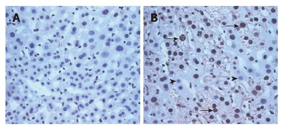
Figure 6.

Double stains for core and 8-OHdG at an age of 2 mo for LAP-tTA transgenics (A) and DTM (B). The density of 8-OHdG was significantly higher in the DTM (B, representatively dark nuclei, arrows, 65% ± 2.4%) than in the LAP-tTA transgenics (2% ± 0.1%). Most 8-OHdG positive cells exhibited robust expression of HCV core. The 8-OHdG negative nuclei were indicated by arrow heads (B).
