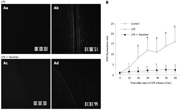
Figure 5.
A: Representative images of the changes in fluorescence intensity of the H2O2-sensitive probe DHR in the rat mesentery venular wall of LPS group (top) and LPS plus compound Danshen injection group (bottom) at 0 min (a and c) and 60 min (b and d) after LPS infusion. The DHR fluorescence on the venular wall was diminished by treatment with compound Danshen injection at 60 min (d) after LPS infusion; B: Time course of changes in DHR fluorescence ratio on the venular walls in different groups. Control: Control group; LPS: LPS group; LPS + Danshen: LPS plus compound Danshen injection group. Data are mean ± SD from 6 rats. aP < 0.05 vs 0 min; cP < 0.05 vs LPS group.

