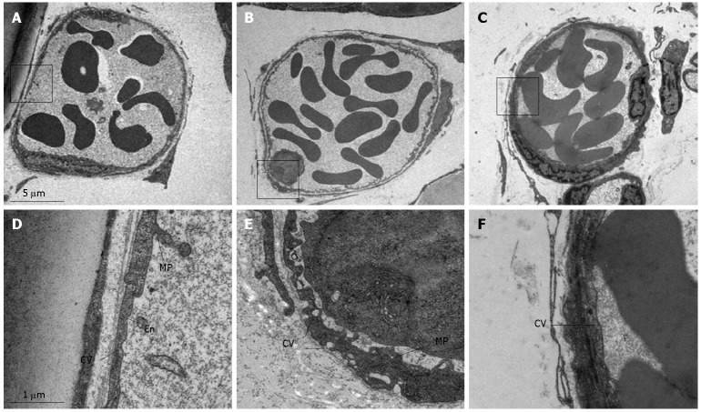
Figure 8.
Representative electron micrographs of post-capillary venules of rat mesentery in the control group (A and D), LPS infusion group (B and E) and LPS plus compound Danshen injection group (C and F). En: Endothelial cell; MP: Microprojection; CV: Cytoplasmic vesicle; L: Leukocyte. Magnification: A: 4000 x; B: 4000 x; C: 4000 x; D: 24 000 x; E: 24000 x; F: 24 000 x.

