
Figure 2.
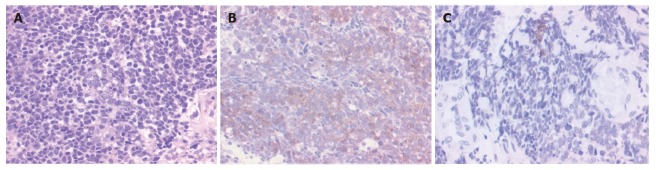
A: The tumor consists of cells with little cytoplasm and round nuclei; HE (hematoxylin-eosin) stain (× 200); B: The tumor tissue diffusely expresses NSE; NSE (neuron-specific enolase) stain (× 200); C: The tumor tissue shows weakly positive expression; Keratin-wide stain (× 200).