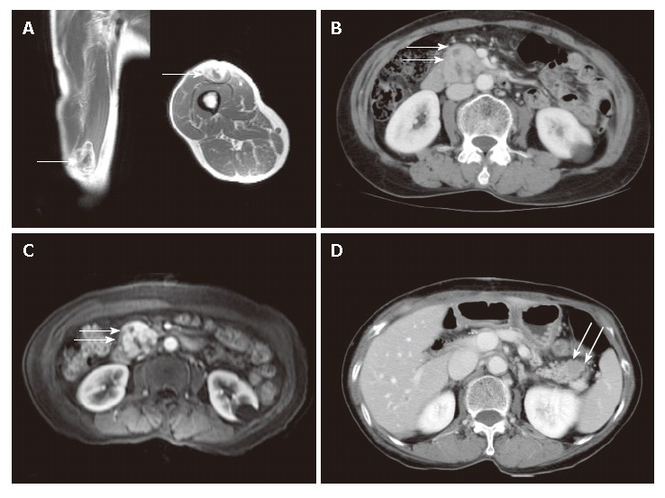
Figure 1.

Radiological findings. A: Gadolinium-enhanced T1 weighted image showing a soft tissue tumor in the muscle of the anterior thigh; B: Contrast-enhanced abdominal CT scan showing a heterogeneously enhanced mass at the head of the pancreas; C: Gadolinium-enhanced T1 weighted image showing a well-enhanced mass with necrotic foci at the head of the pancreas; D: Follow-up CT obtained after 10 mo showing a low attenuated mass in the tail of the pancreas.
