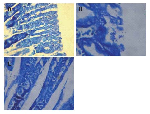
Figure 1.

H pylori stained with Giemsa in gastric tissue of mice. No H pylori found in H pylori antigen + chi-particles group (A) , and lots of H pylori found on surface of gastric mucosa (B) and in gastric foveola (C) of control group (Giemsa dyeing × 400).
