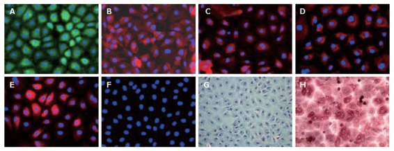
Figure 3.

Immunofluorescence characterization of LECs in primary culture (× 200). Immunofluorescence was visualized using anti-rabbit FITC (green) and anti-mouse Cy3 (rouge). The nuclei, stained by DAPI, appeared in blue. A: The expression of AFP, fetal liver marker, reflected their immature status; LECs expressed both hepatocytic (B: ALB; C: CK-18) and biliary markers (D: CK-19; E: CK-7); F: LECs did not express oval cell marker OV-6; G, H: The representative photographs at phase contrast microscopy after PAS staining. LECs in the primary culture were not able to store glycogen (G) as compared to human hepatocytes (H). The nuclei were stained with hematoxylin.
