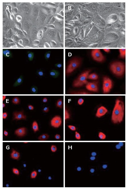
Figure 5.

Characterization of LECs at the 5th passage (× 200). The phase contrast microscopy photographs of LECs at the 5th passage showing an increased size (A and B). Using immunofluorescence, in parallel to morphological changes, LECs lost the expression of AFP (C) and CK-7 (G) but maintained the expression of ALB (D), CK-18 (E) and CK-19 (F). LECs remained mostly negative for oval cell marker OV-6 (H).
