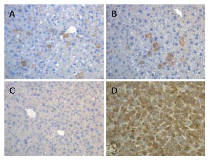
Figure 6.

Immunohistochemical analysis of LECs in the liver of transplanted SCID mice. Foci or isolated cells stained for human Alb (brown) were detected around centrolobular vein (A) and portal area (B) of transplanted SCID mice (2 hepatectomized and 1 normal SCID mice; C: Human Alb cells were not detected in control mice; D: All of cells in human liver sections were stained for human Alb.
