Figure 3.

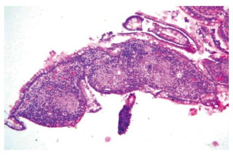
Histological appearance of the mucosal biopsies obtained from the lesion shown in Figure 1. Note the presence of noncaseating granulomas (HE × 40).

Histological appearance of the mucosal biopsies obtained from the lesion shown in Figure 1. Note the presence of noncaseating granulomas (HE × 40).