
Figure 1.
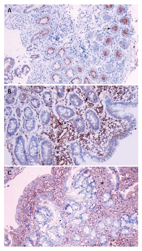
Immunohistochemistry analysis in celiac patients. A: Immunohisto-chemical staining for HLA-G in the Lieberkühn crypts; B: Immunohistochemical staining for IL-10 at the lamina propria level; C: Immunoreaction for TGF-beta in areas of infiltrated inflammatory cells. Arrows and asterisks: cells showing immunoreactivity (x 200).